
Science Topics – 142
The myosin in the sarcomere inside the myocardium repeatedly contracts and relaxes as it swings. In an in vitro test using rat cardiomyocytes, we found that when the muscles that repeat this contraction and relaxation were warmed to a temperature similar to body temperature, they moved with finer contraction and relaxation oscillations. Was named Hyperthermal Sarcomeric Oscillations (HSOs) in the sense of oscillation caused by heat. Furthermore, it was discovered that the small oscillations oscillate in the same period regardless of changes in calcium concentration, that is, there is rhythm homeostasis (CRH). We also built a mathematical model that reproduces that characteristic.
The contraction of the heart is controlled by the change in calcium concentration. However, despite the slow decrease in calcium concentration in cardiomyocytes at the end of systole, the heart rapidly relaxes and dilates, filling with new blood. Until now, I have not understood why this seemingly contradictory phenomenon occurs. This contradiction disappears if we consider that the rapid extension seen in thermal sarcomere vibrations occurs during diastole of the heart. Furthermore, during diastole, ATP consumption is reduced due to the reverse reaction, and it is expected that the heartbeat will be efficient from the viewpoint of energy consumption.
Mechanism of Contraction Rhythm Homeostasis for Hyperthermal Sarcomeric Oscillations of Neonatal Cardiomyocytes. Seine A. Shintani, Takumi Washio, Hideo Higuchi. Scientific Reports: 77443 (2020).
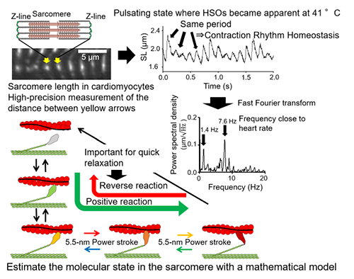
Homeostasis of sarcomere contraction rhythm (CRH) that appears when cardiomyocytes are warmed. Sarcomere is a contraction unit of cardiomyocytes, and the Z-line that divides the sarcomere was visualized with the green fluorescent protein GFP and the distance between the Z-lines was measured (upper left figure). When warmed to 41 °C, sarcomeric oscillation was induced (upper right figure), and the oscillation frequencies were mainly 7.6 and 1.4 Hz (center right figure). It was clarified that the amplitude of the oscillation at 7.6 Hz and the contraction / extension time change greatly, but the period including the contraction and extension time is kept constant (upper right figure). The state transition relationship of myosin in a mathematical model that reproduces this oscillation (lower left figure).
1. Department of Biomedical Sciences, College of Life and Health Sciences, Chubu University
2. Department of Physics, Graduate School of ScienceThe University of Tokyo