
Science Topics - 87
We are able to detect slight changes in the musical rhythm. Recent studies suggest that the cerebellum and basal ganglia are involved in temporal processing for non-motor cognitive functions (e.g. rhythm perception and temporal judgment; Fig. B). Here we show that neurons in the cerebellar dentate nucleus encode the interstimulus interval of isochronous rhythm and play a crucial role in predicting the timing of the next stimulus.
We trained monkeys to detect a single omission of isochronous repetitive stimuli (Fig. A). We found that the dentate neurons responded to each stimulus and gradually elevated the response as the repetition progressed, opposed to the sensory adaptation. The magnitude of the response positively correlated with the interstimulus interval (Fig. C). Because inactivation of the recording sites delayed the detection of stimulus omission, these signals might be necessary for the prediction of stimulus timing. This study revealed the underlying mechanisms of rhythm perception at single-neuron level. Our findings will advance the understanding of the cerebellar disorder and encourage future clinical techniques for the diagnosis and the evaluation of treatment.
Ohmae S, Uematsu A, Tanaka M. “Temporally specific sensory signals for the detection of stimulus omission in the primate deep cerebellar nuclei.” J Neurosci 2013; Sep 25
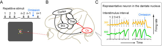
(A) Missing oddball task. During monkeys looked at the fixation point (red dot) on the monitor, visual stimuli (white square) were presented repeatedly at a fixed interval. Tones were also presented simultaneously. As monkeys reported the omission of the audiovisual stimulus by making a saccadic eye movement, drops of juice were given as a reward. (B) Neural circuitry related to the temporal processing. Cb, Cerebellum; Thal, Thalamus; BG, Basal ganglia; Cortex, Cerebral cortex. (C) Example of a neuron recorded from the cerebellar dentate nucleus. In the left and right panels, data were aligned with the first stimulus and the omission of the stimulus, respectively. Vertical dashed line indicates the timing of each repetitive stimulus. Note that the response was greater as the repetition progressed and the interstimulus interval was longer.
1Department of Physiology, Hokkaido University School of Medicine (2Department of Psychology, University of Pennsylvania)