
Science Topics - 96
Mammals exhibit a variety of physiological responses to psychological stress, such as increases in body temperature, heart rate and blood pressure. These stress responses have a biological significance by increasing physical performances to survive a crisis of life, such as confrontation with enemies. In the modern society, however, many people suffer from stress disorders caused by excessive psychological stressors. For example, psychogenic fever caused by chronic psychological stress is a difficult psychosomatic symptom due to its resistance to antipyretics.
We sought to determine the central circuit mechanism that drives stress-induced hyperthermia. Rats given social defeat stress, a sociopsychological stress model, exhibited increases in sympathetic heat production by brown adipose tissue and in body temperature. Drug nanoinjections into the brain revealed that activation of neurons in the dorsomedial hypothalamus (DMH) and rostral medullary raphe (rMR) mediates the stress-induced responses. We also found that neurons projecting from the DMH to the rMR are activated by social defeat stress. Specific stimulation of those projection neurons using an optogenetic technique elicited increases in brown adipose tissue thermogenesis, heart rate and blood pressure, mimicking stress responses.
These results indicate that the direct pathway from the DMH to the rMR mediate the stress signaling that drives hyperthermia and other sympathetic responses (Figure). We also found a stress-activated pathway from the DMH to the paraventricular hypothalamic nucleus, a neuroendocrine center for stress hormone release (Figure). Our present findings may contribute to future development of therapies for stress disorders including psychogenic fever
Kataoka N, Hioki H, Kaneko T & Nakamura K (2014) Psychological stress activates a dorsomedial hypothalamus–medullary raphe circuit driving brown adipose tissue thermogenesis and hyperthermia. Cell Metabolism 20(2):346-358 (2014).
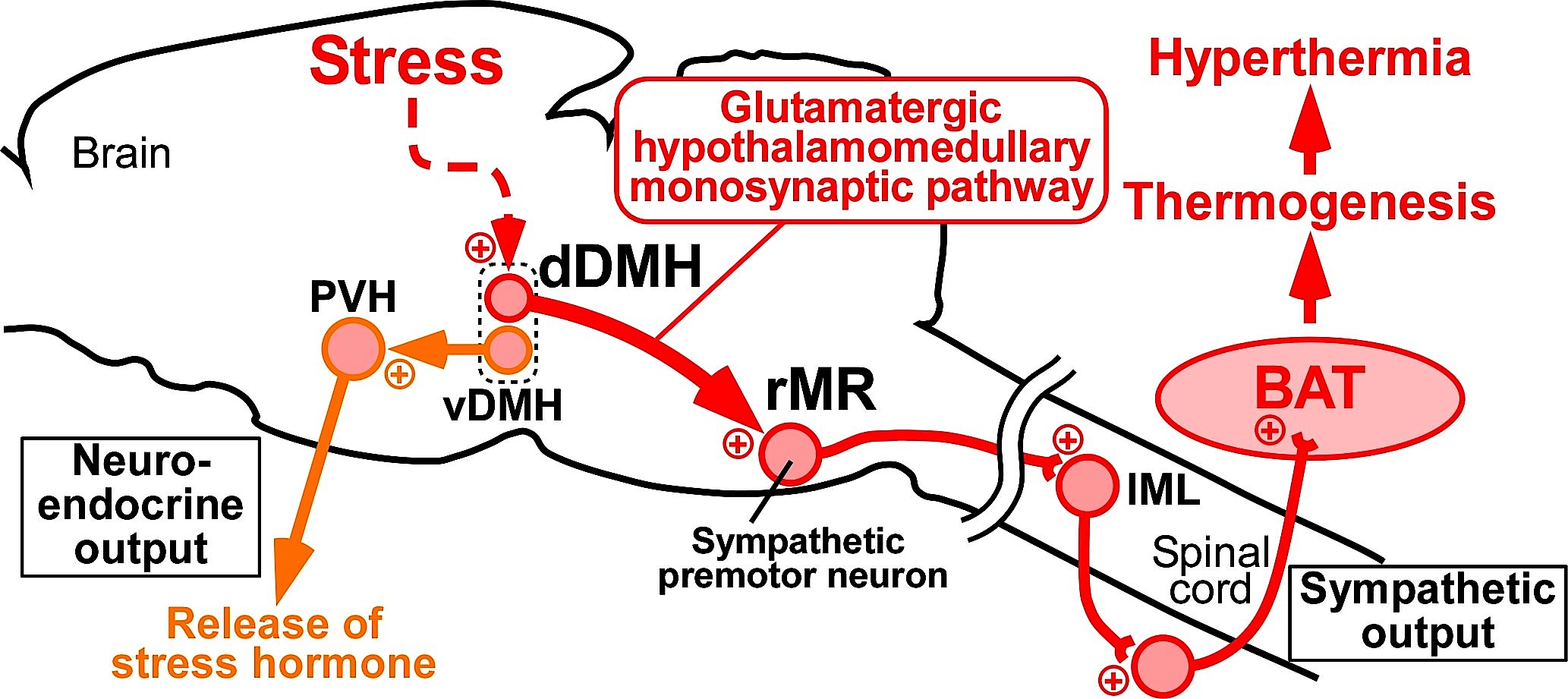
Figure: Schematic central circuits for sympathetic and neuroendocrine stress responses. Forebrain stress signals activate two groups of DMH neurons: neurons in the dorsal part (dDMH) provide a direct glutamatergic input to sympathetic premotor neurons in the rMR to drive brown adipose tissue (BAT) thermogenesis contributing to hyperthermia, and neurons in the ventral part (vDMH) provide a direct input to the paraventricular hypothalamic nucleus (PVH) to drive a neuroendocrine outflow to release stress hormones. Plus signs indicate excitatory neurotransmission. IML, intermediolateral nucleus.
Career-Path Promotion Unit for Young Life Scientists, Kyoto University